

 Slovensky (EUR)
 Česky (CZK)
 Magyar (HUF)
Endothelium: the living lining of blood vessels and its role in circulation
Endothelium: the living lining of blood vessels and its role in circulation
The endothelium is a thin layer of living cells lining the inner surface of blood vessels. It helps regulate vessel dilation, permeability and contact between blood and the vessel wall. This article explains its functions, endothelial dysfunction and links with atherosclerosis. It also distinguishes the endothelium from the deeper layers of an artery and biological mechanisms from effects investigated in research.
The main guide to the whole system is 03 · Heart and blood vessels. Here, we focus on the inner lining of blood vessels.
01 · What is the endothelium and what does it do?
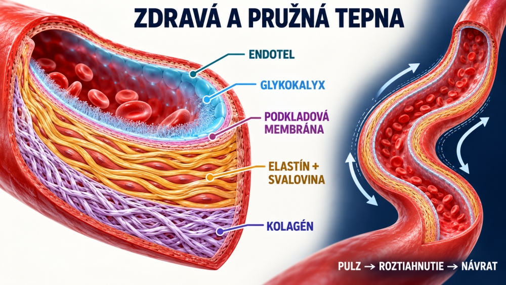
Imagine blood vessels as a road network. The heart maintains blood flow, arteries and veins form the main routes, and the smallest capillaries bring blood close to cells. The endothelium is a thin, living layer of cells lining the inside of blood vessels. It responds to blood flow and helps regulate vessel tone, the permeability of the vessel wall and the interaction between blood and the vessel lining.
A continuous layer of cells and the junctions between them help regulate the barrier between blood and the vessel wall.
A delicate surface layer of proteins and sugar chains. Its components include proteoglycans, heparan sulphate and hyaluronan.
A layer beneath endothelial cells containing type IV collagen, laminin and other components.
Where is elastin? Elastic fibres are mainly found in the deeper walls of larger arteries; they are not the principal structural component of the thin endothelial cell layer. Collagen is also present beneath the endothelium, but its type and role vary between vessel layers.
A delicate layer called the glycocalyxlies on the endothelial surface. It consists of various molecules, including glycosaminoglycans; one component can be hyaluronan, a form of hyaluronic acid. It helps create an interface between blood and vessel cells. The endothelium also produces nitric oxide (NO), which contributes to the regulation of blood vessel dilation.
The endothelium also helps regulate blood clotting and inflammatory processes. Its function varies with the type of vessel and tissue; a single diagram therefore cannot show every form of the vascular lining.
02 · Endothelial dysfunction: when the lining does not work properly
Long-term high blood pressure, elevated LDL cholesterol, smoking or diabetes change the environment in which the endothelium operates. Inflammatory processes, age and blood flow patterns also matter, particularly at arterial branches. Reduced NO availability and impaired protective endothelial function are associated with endothelial dysfunction.
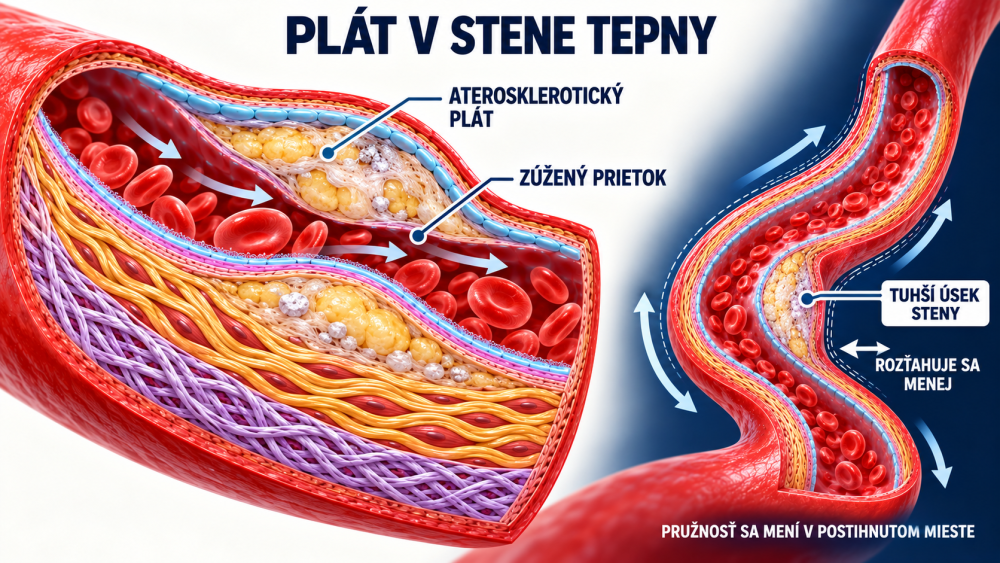
A “stiff endothelium” and a “stiff blood vessel” are different concepts. Arterial stiffness involves the whole arterial wall: elastic fibres, collagen, smooth muscle, any calcification and pressure. The endothelium can contribute to the regulation of vessel tone, but it does not constitute the entire elastic wall.
Related risk factors are explained in 08 · Blood pressure and 09 · Blood sugar. Blood pressure or glucose readings alone are not direct measurements of endothelial function.
03 · Collagen, elastin and hyaluronic acid: what are their roles?
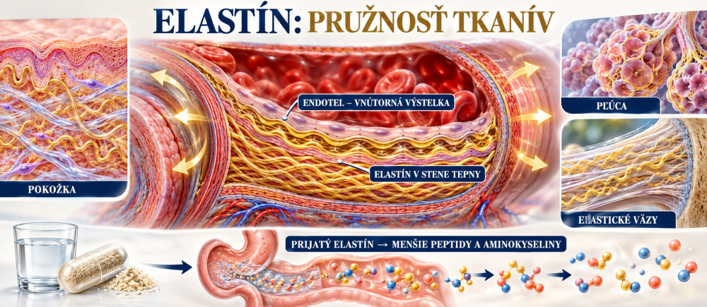
Elastic fibres in the arterial wall allow it to stretch and recoil with each pulse.
Contributes to the strength of the vessel wall. The proportions and arrangement of fibres matter.
One component of the environment and glycocalyx around the endothelium; it is not a “lubricant” that coats vessels after it is swallowed.
With ageing, elastic fibres can change and fragment; collagen and calcification also contribute to the stiffness of large arteries. Ingested collagen and elastin do not attach to the arterial wall as ready-made new fibres. Proteins undergo digestion, producing amino acids and smaller peptides. The body can use these, but restoration of a particular blood vessel cannot be promised after taking a supplement.
The journey of nutrients from digestion to their use in the body is discussed in 06 · From intake to use in the body.
04 · Endothelium and atherosclerosis: how plaque forms
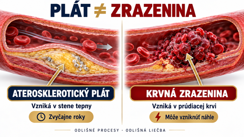
Not exactly. Atherosclerotic plaque develops within the arterial wall and evolves over years. It can contain cholesterol-rich lipids, inflammatory cells, connective tissue and calcium. Its composition varies: some plaques are more lipid-rich, others more fibrous or calcified. Plaque can narrow the space available for blood flow; another danger is disruption of its surface and the formation of a blood clot.
The pipe analogy helps us imagine narrowing, but it does not replace biology. Plaque does not usually “soak off” the wall like a layer of grease. Treatment of risk factors can slow its growth and reduce complications. In clinical studies, intensive LDL-lowering treatment has also produced measurable reductions in plaque volumein some patients; this does not mean that all plaques disappear immediately. For severe narrowing, a doctor may recommend a vascular procedure depending on the circumstances.
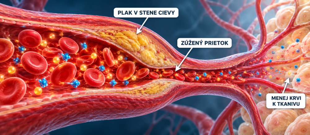
Plaque formation and the role of cholesterol are explained in more detail in 07 · Atherosclerosis and cholesterol.
05 · A capillary can be narrower than a red blood cell
At rest, a red blood cell is a biconcave disc with a diameter of approximately 7–8 micrometres (µm). Capillaries do not have a uniform diameter: in many tissues they are in the range of 4–10 µm, while the narrowest sections may be approximately 3–5 µm. One micrometre is one thousandth of a millimetre.
red blood cell diameter at rest
narrowest capillary sections – approximate values
If a red blood cell is 8 µm across and a narrow capillary section is 4 µm across, the cell is approximately twice as wide at rest as that section. It passes through because it can deform and then change shape again. It does not pass through by losing mass or by requiring the vessel wall to “open” to eight micrometres. Red blood cells mainly carry oxygen; many dissolved nutrients travel in blood plasma and pass through capillary walls into surrounding tissue.
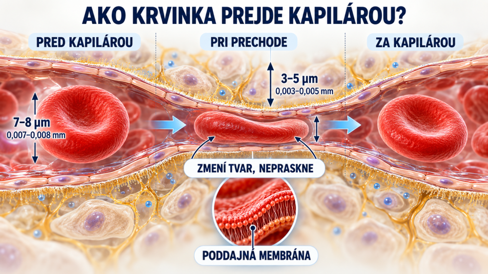
For more on flow through the smallest blood vessels, see 02 · Blood viscosity and microcirculation.
06 · Should blood be as thin as possible?
No. Healthy circulation does not require blood to be “as thin as possible”. Flow properties depend on several factors: the number and behaviour of blood cells, their deformability, plasma, vessel diameter, pressure and local flow. Blood clotting is a separate topic: it is needed to stop bleeding, but under certain circumstances it can contribute to a dangerous clot.
Drinking water supports normal hydration, but it does not “dissolve plaque”. Anticoagulant and antiplatelet medicines should only be taken as prescribed; attempting to “thin the blood” on your own can be dangerous.
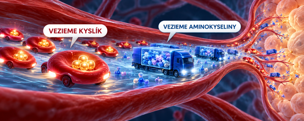
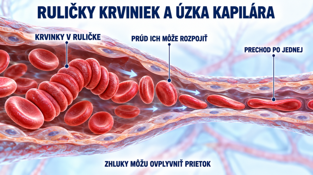
The difference between a blood-drop image and a laboratory examination is explained in 01 · Blood under the microscope. For the capabilities and limitations of individual measurements, see 15 · Heart and circulation measurements.
07 · What nutrients do the endothelium and vessel wall need?
The body needs sufficient energy, protein and ordinary micronutrients to maintain cells and tissues. Vitamin C contributes to normal collagen formation for the normal function of blood vessels – this is an authorised EU health claim when the food meets its conditions of use. Collagen is part of the vessel wall, but ingested collagen is not directed exclusively to it.
Elastin and hyaluronan also have biological roles in the vascular system. However, this does not meanthat taking elastin or hyaluronic acid will restore damaged endothelium, remove calcification or shrink plaque. For most people, a varied diet with enough vegetables, fruit, pulses, wholegrains and suitable protein sources matters more than searching for one “vitamin for the endothelium”.
How does the endothelium repair itself?
Following minor damage, neighbouring endothelial cells can migrate and multiply, form a continuous layer again and restore their junctions. The body also continually produces glycocalyx components. Repair capacity depends on health status and whether harmful influences on the vessels persist.
Normal cell function requires oxygen, nutrients and ordinary metabolism. In practical terms, the priority is to reduce repeated damage: stop smoking, treat high blood pressure, elevated LDL and diabetes according to medical advice, and exercise regularly at a suitable level. Nutrient deficiencies should be addressed individually; there is no pill that gives the entire endothelium a “new lining”.
B vitamins and other nutrients have their usual roles in metabolism. If a deficiency is suspected, assess its specific cause and the test results; no universal cocktail can replace the management of blood pressure, LDL or blood sugar.
09 · How does science investigate light and blood vessels?
Scientists are exploring new possibilities and studying photobiomodulation – the use of precisely controlled red or near-infrared light. Different wavelengths, doses, application sites and methods are studied; findings cannot automatically be transferred from one method to another.
In 2021, Yin and colleagues studied 635 nm light in cells and by irradiating the chest and abdomen of mice with atherosclerosis. After ten weeks, the irradiated mice had a smaller plaque area; the authors also investigated cholesterol efflux from macrophages. This was not a human study or evidence of plaque dissolution. [3]
Weihrauch and colleagues observed effects of 670 nm light on blood vessels and signalling mechanisms associated with nitric oxide. This work investigated vessel dilation, not the removal of human plaques. [4]
In a small randomised study, the doses used did not produce a significant improvement in measured endothelial function compared with placebo. This finding cannot be applied to every wavelength either. [5]
What can we say today? Some experiments suggest biological effects of light on vascular cells and on atherosclerosis in mice.
These experiments do not establish that an ordinary home device restores damaged endothelium or removes human plaques. Findings must be assessed according to the specific method, dose and study population; they do not replace standard treatment.
For more on methods and limitations in light research, see 13 · Laser blood irradiation and 14 · Cells and photobiomodulation.
10 · Frequently asked questions
Is the endothelium the same as the whole vessel wall?
No. It is the wall's inner cellular lining. Deeper layers of large arteries contain smooth muscle, elastic fibres and collagen.
Is every older artery “blocked with fat”?
No. Arterial stiffness and atherosclerotic plaque are related but distinct processes. Plaque varies in size and composition, and not every stiffer artery has significant narrowing.
Can plaque shrink?
In some patients, intensive LDL-lowering treatment has produced measurable reductions in plaque volume. The main aim is to reduce the risk of heart attack and stroke; there is no simple home method for “dissolving deposits in blood vessels”.
How does a blood cell pass through a narrower capillary?
It is flexible and changes shape as it passes through. A diameter of 7–8 µm describes the cell at rest, not a rigid spherical load.
Is collagen or hyaluronic acid enough to restore the endothelium?
It has not been established that a supplement alone restores damaged endothelium or removes plaque. The appropriate approach depends on risk factors and health status.
11 · Related articles and scientific sources
Scientific sources – expand
- [1] Endothelial structure: glycocalyx and basement membrane.
- [2] Endothelial repair: cell migration, proliferation and junctions.
- [3] Yin et al. (2021): 635 nm photobiomodulation and plaques in mice.
- [4] Weihrauch et al. (2021): 670 nm and experimental vessel dilation.
- [5] Hauck et al. (2023): an 810 nm trial in healthy people with no improvement over placebo.
- [6] NHLBI/NIH: atherosclerosis, plaque composition and risk factors.
- [7] NHLBI/NIH: atherosclerosis treatment.
- [8] ASTEROID: changes in plaque volume with intensive statin treatment.
- [9] GLAGOV: a randomised study of coronary plaque changes.
- [10] Elastin, arterial mechanics and elasticity.
- [11] The endothelial glycocalyx and vascular function.
- [12] Blood cell deformation under flow: a review of mechanical forces.
- [13] EU Regulation 432/2012: the authorised claim for vitamin C and blood vessels.
- [14] Red blood cell biomechanics: dimensions and passage through narrow capillaries.
Educational information: This article is educational and does not provide a diagnosis or treatment. If you take medicines for blood pressure, cholesterol or blood clotting, do not change them without consulting your doctor. For sudden chest pain, speech difficulties or weakness on one side of the body, call 112 in the EU or your local emergency number.