

 Slovensky (EUR)
 Česky (CZK)
 Magyar (HUF)
 Slovenščina (EUR)
 Deutsch (EUR)


02. Blood under the microscope: what a drop of blood can and cannot show

KINGRAY ACADEMY · ARTICLE 02
Blood under the microscope: what a drop of blood can and cannot show
What microscopic observation of blood can tell us
For a non-specialist, a microscopic image of blood is primarily educational. In the hands of a trained professional, with appropriate sample preparation and validated haematological methods, microscopy is an important tool that complements a complete blood count and other laboratory tests.
At KINGRAY, we use both brightfield and darkfield microscopy. These allow us to observe the appearance and arrangement of blood cells, aggregate formation and changes in the image over time. These observations describe the particular sample being examined; on their own, they do not demonstrate that the blood behaves in the same way within the circulation.
Proper interpretation requires knowledge of blood cells, the effects of plasma proteins and changes that occur after collection. The quality of the preparation, the timing of observation, a consistent procedure and the observer’s practical experience all matter.
A trained professional distinguishes between what is visible in the sample, possible explanations for a finding and what needs to be confirmed through further testing. Diagnostic conclusions drawn from a standardised blood smear cannot automatically be applied to a fresh, unstained drop. Using brightfield or darkfield illumination alone does not turn observation of a fresh drop into a validated diagnostic test.
Under a microscope, a drop of blood reveals blood cells and their arrangement in the sample. The appearance of a fresh drop alone cannot establish a diagnosis or determine cholesterol, blood glucose or a numerical blood viscosity value.
We explain the components of blood, the appearance of red and white blood cells, the meaning of rouleaux formation and how fibrin forms. We also distinguish educational observation of a fresh drop from a laboratory blood smear and a complete blood count.
01 · What is blood made of?
Blood consists of plasma, red blood cells, white blood cells and platelets. Each component has a different role, so they are not all assessed in the same way under a microscope. [1]
Blood components: their functions and relevance to microscopic observation
| Blood component | Main role | What matters when observing a sample |
|---|---|---|
| Blood plasma | Main role: A fluid containing water, proteins and other dissolved substances; it transports nutrients, hormones and metabolic waste products. | When observing: It forms the fluid surrounding the cells. The concentrations of its individual components cannot be determined from their appearance. |
| Red blood cells – erythrocytes | Main role: They use haemoglobin to carry oxygen from the lungs to tissues. | When observing: We can observe their outline and arrangement. Diagnostic assessment of their morphology requires a properly prepared blood smear. |
| White blood cells – leukocytes | Main role: They are part of the immune system and help defend the body. | When observing: Their types are distinguished through standardised laboratory testing; appropriate staining of a blood smear reveals further details. |
| Platelets – thrombocytes | Main role: Small cell fragments that help stop bleeding. | When observing: They are smaller than blood cells. Neither their laboratory count nor their function can be determined simply by looking at a fresh drop. |
02 · What can we see in a drop of blood under a microscope?
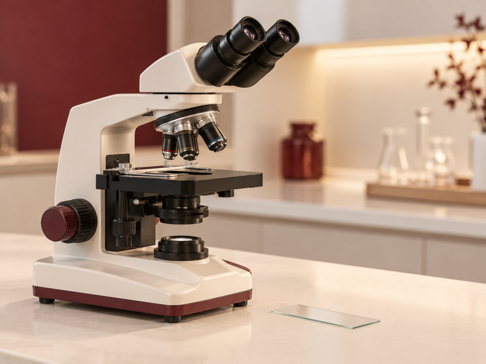
A microscope brings the cellular components of blood into view. What we can distinguish depends on sample preparation, lighting and magnification.
When we examine a fresh drop of blood under a microscope, some of the most prominent structures are red blood cells – erythrocytes.
Red blood cells contain haemoglobin, and their main role is to carry oxygen from the lungs to tissues and contribute to the transport of carbon dioxide back towards the lungs.
What can we observe? A microscope allows us to observe cells and other structures in a sample. However, the appearance of a fresh drop is not a complete blood count. A standardised laboratory blood smear has diagnostic uses alongside other tests. [2]
03 · Red blood cells, rouleaux and fibrin
When examining a sample under a microscope, we may see red blood cells in different arrangements, as well as structures that form as the sample clots.
Individual red blood cells
A properly prepared sample can show individual, clearly distinguishable red blood cells.
A typical red blood cell has the shape of a biconcave disc – slightly indented on both sides. This shape and its flexibility help it transport oxygen and pass through very small blood vessels.
Rouleaux formation – red blood cells in “stacks of coins”
Sometimes red blood cells lie close together, creating an appearance resembling coins stacked on top of one another.
This phenomenon is called rouleaux. Its formation can be influenced by the plasma environment, the concentration of certain plasma proteins, sample preparation and the time of observation. The finding alone is not evidence of a specific disease. Rouleaux is not the same as a blood clot. [4]
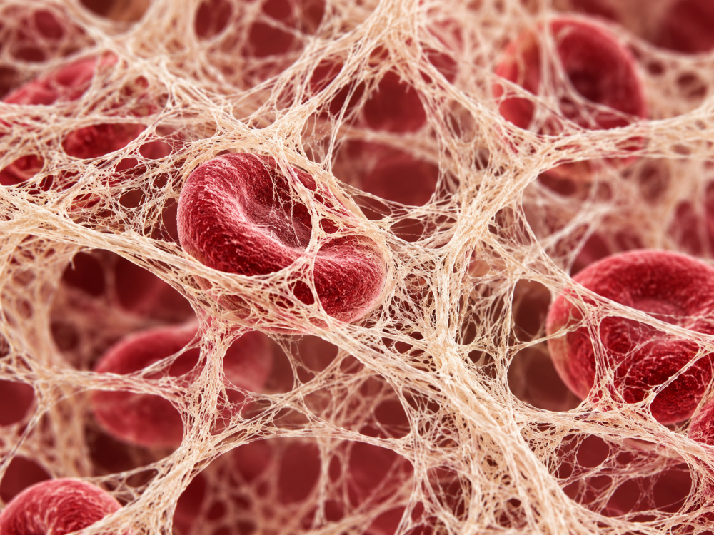
Fibrin and clotting
Fibrin is part of the natural blood clotting process. It forms a fibrous network that helps stabilise a developing clot.
This network can trap platelets and blood cells.
Clotting of a collected drop outside the body does not, by itself, demonstrate a clot in the person’s blood vessels. [5]
Important: The appearance of a single drop of blood cannot be used to diagnose a disease on its own. Sample preparation and the time since collection can also affect the image.
04 · What does a typical red blood cell look like?
Typical appearance
- A regular, round outline.
- A paler centre in a conventional microscopic blood smear.
- A biconcave shape.
- Flexibility that matters when passing through small vessels; a static image does not measure it.
- The ability to deform when passing through capillaries.
What can we observe?
Under a microscope, we may see individual red blood cells, cells lying close together, different cell arrangements or structures forming as the sample clots.
However, the appearance of one drop alone is not enough to establish a diagnosis.
Interpret with care. A microscopic image should be described in terms of what is visually observed in the sample. Specific diseases or laboratory values cannot be determined from it without the appropriate laboratory tests.
05 · Blood cells as cars: a visual analogy
Imagine red blood cells as small vehicles constantly travelling through the body.
Separate blood cells
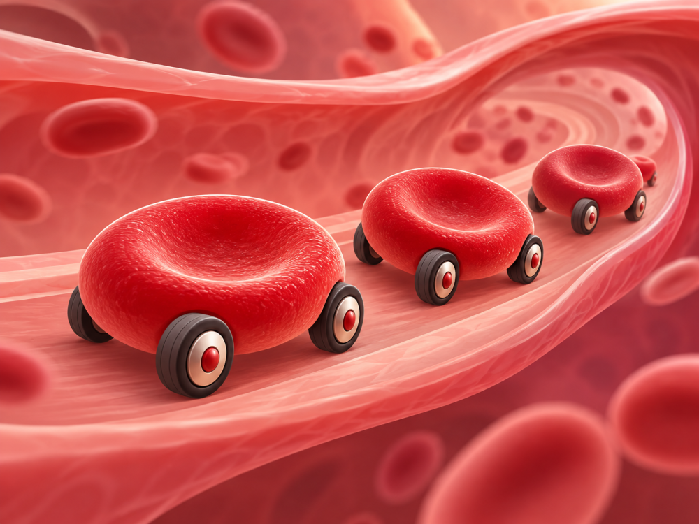
Individual blood cells can be clearly distinguished in a properly prepared sample.
Rouleaux
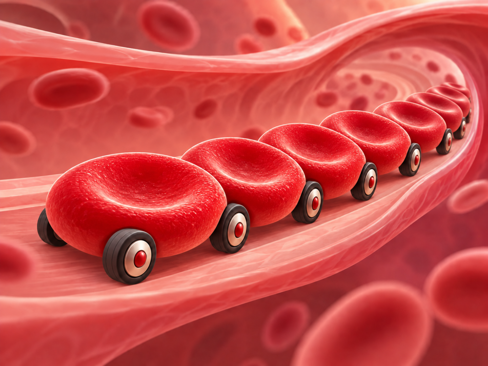
Blood cells may form an arrangement resembling cars lined up closely together. This is neither a traffic accident nor a literal representation of blood flow.
A clot
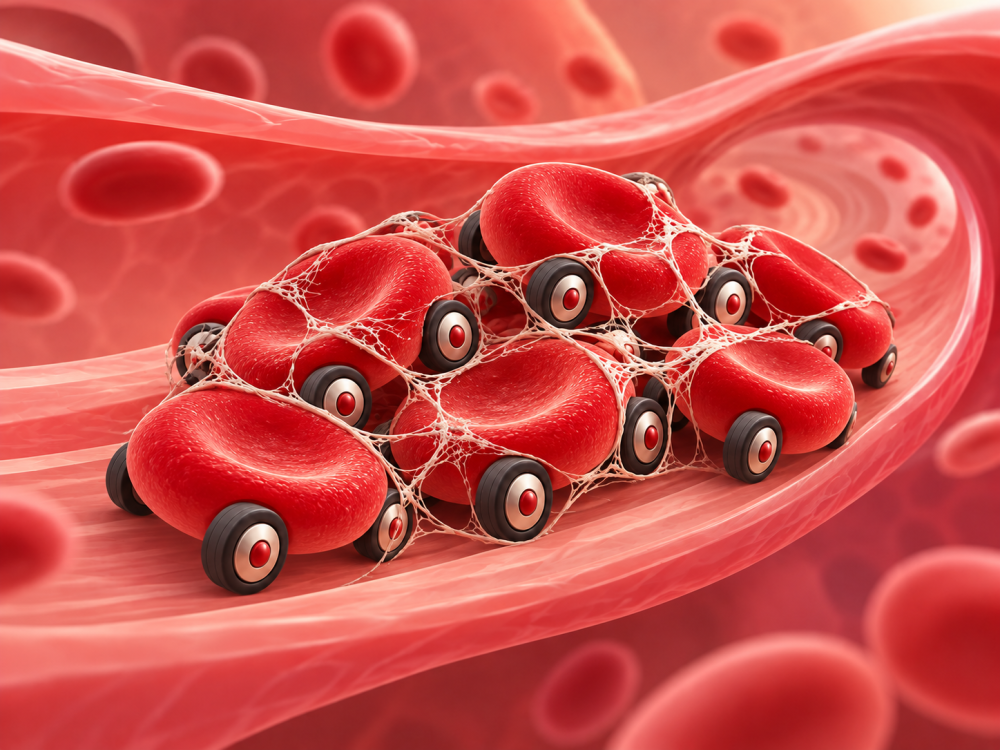
Fibrin forms a network that helps stabilise a blood clot.
The analogy is only an aid to understanding. A microscopic image of blood is not a direct view of how blood flows through vessels in a living body.
06 · Fibrinogen and fibrin: what is the difference?
Fibrinogen is a soluble protein in blood plasma. When clotting is activated, it is converted into fibrin, which forms a fibrous network that stabilises a developing clot.
The key point for this article: We may observe structures forming as a sample clots, but their appearance does not determine a laboratory fibrinogen concentration or a numerical blood viscosity value.
Want to understand how fibrinogen relates to viscosity?
Blood viscosity and microcirculation →
07 · A fresh drop and laboratory tests
The difference between observation and laboratory tests
A blood smear examines the appearance of cells; a complete blood count provides numerical data. Neither automatically measures cholesterol, glucose or fibrinogen concentration – these require the relevant laboratory tests. Results are assessed alongside symptoms and the person’s health status. [2] [3]
Which measurement answers which question? Continue to article 15 · Heart and blood circulation measurements, which compares blood pressure, pulse, ECG, HRV and microscopic observation.
08 · What affects the sample, and what can it not tell us?
Microscopic observation of a fresh drop of blood at KINGRAY has an educational purpose.

It is not a complete blood count and is not used to diagnose, treat or rule out disease.
The appearance of a specimen may be affected by:
- The collection method.
- Sample preparation.
- Pressure from the coverslip.
- Temperature.
- Time since collection.
What can we not determine from a drop of blood? The visual appearance of a drop cannot determine a numerical blood viscosity value, laboratory parameters or a specific medical diagnosis.
Viscosity cannot be read from a photograph of a drop of blood
To learn what actually affects it and how it relates to haematocrit, plasma and red blood cells, continue to the next article.
Explore blood rheology →
09 · Frequently asked questions about blood under the microscope
Short answers to common questions about microscopic observation of a fresh drop of blood.
What are live blood observation and darkfield microscopy?
“Live blood” refers to observing a fresh, unstained blood sample. Darkfield microscopy shows light-scattering structures as bright objects against a dark background. It is an imaging technique; it does not, by itself, establish that an interpretation can reliably diagnose disease.
Can white blood cells under a microscope reveal immune health?
White blood cells (leukocytes) are part of the immune system. Their movement or lack of movement in a fresh drop cannot reliably indicate the “strength” of immunity. A complete blood count with differential measures the number and types of white blood cells; results are assessed alongside symptoms and other findings. [3]
Are echinocytes evidence of toxins in the body?
No. Echinocytes, also called burr cells, are red blood cells with short, fairly evenly spaced projections. This appearance can also result from sample preparation or storage. The image alone does not demonstrate “toxins” or an antioxidant deficiency. [6]
What is microscopic observation of a fresh drop of blood?
It involves examining a fresh drop of blood under a microscope to visually observe blood cells and other structures in the sample. It has an educational purpose and does not replace laboratory testing.
What do red blood cells look like?
Red blood cells have a typical biconcave shape. In a conventional microscopic blood smear, they appear as round cells with a paler centre.
What does it mean when red blood cells form “stacks of coins”?
One possible appearance is rouleaux – an arrangement of red blood cells resembling coins stacked on top of one another. Its formation may relate to the plasma environment and concentrations of certain plasma proteins. Sample preparation and the time of observation also affect the image; it does not identify a specific disease on its own.
What is fibrin?
Fibrin forms during blood clotting. It creates a fibrous network that helps stabilise a developing blood clot.
What is the difference between fibrinogen and fibrin?
Fibrinogen is a soluble protein naturally present in blood plasma. When clotting is activated, fibrinogen is converted into fibrin, which forms a fibrous network.
Can a drop of blood show how “thick” the blood is?
Visual observation of a drop of blood cannot determine a numerical blood viscosity value. Viscosity is a physical quantity that requires an appropriate measurement method.
Can a microscopic image establish a diagnosis?
Looking at a fresh drop alone cannot. However, a standardised laboratory blood smear has diagnostic uses. Microscopic observation of a fresh drop at KINGRAY has an educational purpose. It does not replace laboratory testing, medical diagnosis or professional clinical assessment.
Can sample preparation affect its appearance?
Yes. The appearance of a specimen may be affected by the collection method, sample preparation, coverslip pressure, temperature or the time elapsed since collection.
Does fibrin in a collected drop mean there is a clot in my blood vessels?
No. A collected sample may start to clot outside the body. This observation alone neither proves thrombosis nor rules out disease. A suspected health problem requires professional assessment.
10 · Sources
Sources and further reading – expand
Sources on blood components, laboratory morphology and the limits of interpretation:
- [1] American Society of Hematology – Blood Basics: blood components and their basic functions.
- [2] NIH / MedlinePlus – Blood Smear: preparation and uses of a laboratory blood smear.
- [3] NIH / MedlinePlus – Complete Blood Count: information provided by a complete blood count.
- [4] American Society of Hematology – Rouleaux formation: micrographs showing red blood cell arrangements.
- [5] NIH / MedlinePlus – Blood clot formation: the roles of platelets and fibrin in clotting.
- [6] American Society of Hematology – Burr Cells or Echinocytes: the appearance of echinocytes and possible artefacts caused by sample preparation or storage.
- Reliability of Enderlein’s darkfield analysis of live blood (2006): research into the reliability of a specific method of assessing fresh blood; it does not evaluate all laboratory microscopy methods.
Know your body.
Start with your heart.