

 Slovensky (EUR)
 Česky (CZK)
 Magyar (HUF)
 Slovenščina (EUR)
 Deutsch (EUR)


09. Chronic Inflammation and Ageing: What Does Inflammaging Mean?

Chronic inflammation and ageing: what does inflammaging mean?
Inflammation helps protect the body and repair damaged tissues. When it persists, however, it can impair vascular function, metabolism and regeneration. Healthy longevity therefore depends on what sustains inflammation and on the conditions in which cells function: adequate oxygen, nutrients and energy, functioning microcirculation and the ability to end the defence response once its cause has been removed. CRP, hs-CRP and the erythrocyte sedimentation rate help assess this situation, but each captures a different part of the picture.
Sources for the introduction: [1] [2] [7] [10] [11]
When inflammation helps and when it harms
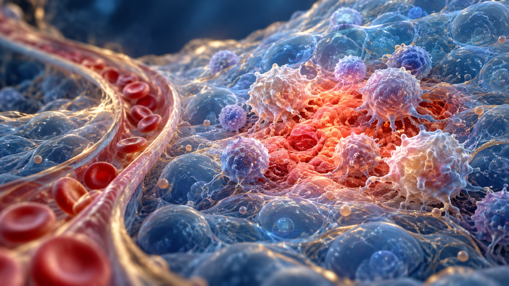
Acute inflammation occurs, for example, during infection or after injury. Defence cells arrive at the affected site, and blood flow and vascular permeability change. This may cause redness, warmth, swelling, pain and temporary loss of function. Once the trigger has been removed, the response should resolve in a controlled way and the tissue should recover.
Chronic inflammation develops when a trigger persists, damage recurs or the mechanisms that end the inflammatory response do not work properly. It need not cause fever or marked pain. It may remain local or affect the whole body. Causes include persistent infections, autoimmune diseases, metabolic disorders, irritants and tissue damage without infection. [7]
Some people develop persistent low-grade inflammatory activity during ageing, known as inflammaging. It is associated with immune changes, accumulated cellular damage, the environment and lifestyle. It is not a separate diagnosis or a process that necessarily follows the same course in everyone. Comparisons between populations show that the relationship between age and inflammatory markers also depends on living conditions. [1] [6] [7]
Where chronic inflammation originates
The intestines and blood vessels can both be important sites of inflammation. Its source cannot automatically be attributed to a single organ in everyone. Depending on symptoms and findings, an assessment considers several possibilities:
- Mouth and teeth. Inflammation of the gums and tooth-supporting tissues, or an untreated dental infection, can place a prolonged burden on the body.
- Intestines. Inflammatory bowel disease, coeliac disease and certain infections activate the immune system. Some conditions also involve disruption of the intestinal barrier and exposure to microbial components. Bloating alone does not identify the cause or extent of inflammation.
- Visceral fat and metabolic disorders. Fat around the internal organs is metabolically active. Excess visceral fat, insulin resistance and metabolic liver disease can promote low-grade inflammation.
- Blood vessel walls. In atherosclerosis, inflammation develops in connection with the retention of apoB-containing lipoprotein particles and the subsequent immune response. Vascular care therefore also addresses blood lipids, blood pressure, blood glucose and smoking.
- Other tissues and diseases. Chronic wounds, certain lung diseases, persistent infections, autoimmune conditions and repeated tissue injury require their own diagnostic and treatment approaches. [5] [7] [9]
Inflammatory mediators from one site can travel in the blood and affect distant organs. The liver, for example, responds by producing more CRP. A systemic inflammatory response can therefore occur without infection spreading throughout the body. Smoking, physical inactivity, sleep disturbances and prolonged metabolic strain may further contribute. [2] [7]
Microcirculation and the conditions for tissue repair
Microcirculation is blood flow through the smallest vessels, where oxygen, nutrients and metabolites are exchanged between blood and tissues. Cellular health depends not only on blood reaching an organ, but also on its distribution within small vessels and the ability of substances to reach the cells.
Blood flow often increases at the start of acute inflammation. Dilated vessels help deliver defence cells and necessary substances. In severe or persistent inflammation, however, increased vascular permeability, swelling and impaired vessel lining can hinder tissue supply. The distance oxygen must travel from a capillary to a cell also increases. Inflammation therefore does not always mean reduced blood flow; its phase, extent and the tissue affected matter. [11]
Ischaemia means inadequate blood supply; hypoxia means insufficient oxygen. Ischaemia can damage cells and trigger inflammation. Under some circumstances, inflammation can further impair blood supply. Oxygen shortage and inadequate removal of metabolites may also alter local pH. This local tissue acidification must be distinguished from systemic blood acidosis; urine pH does not reveal the pH of a particular inflamed tissue. [11]
Reactive oxygen species, chemically active molecules derived from oxygen, contribute to defence and cellular signalling. When their production exceeds the cell’s regulatory and protective capacity, oxidative stress develops and can damage membranes, proteins and DNA. Inflammation, hypoxia and oxidative damage can then reinforce each other. The need to repair and replace damaged cells increases; some cells die and others enter senescence. Regeneration requires suitable conditions and removal of the cause of damage. [3] [7] [11]
How inflammation connects with cellular ageing
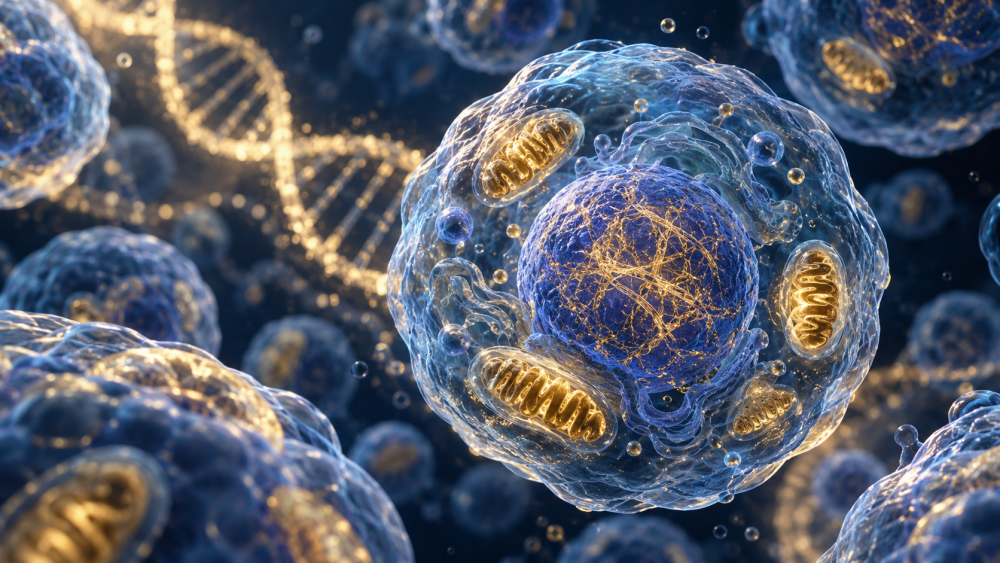
Senescent cells stop dividing but remain metabolically active. Some release a mixture of inflammatory and other signalling substances called the SASP. These signals can affect nearby cells and, when they accumulate over time, contribute to an unfavourable tissue environment. Senescence also has protective roles, such as limiting the proliferation of damaged cells. [3]
Another mechanism under investigation is the NLRP3 inflammasome, part of innate immunity that responds to certain danger and damage signals. Mouse experiments have demonstrated its involvement in inflammation and age-related functional decline. Such findings help explain a mechanism but do not, on their own, establish a treatment for healthy people. [4]
Chronic inflammation and cellular ageing can therefore reinforce one another. Their significance is assessed alongside organ function, metabolic health, physical performance and specific diseases. A single blood test cannot capture the rate of ageing of the entire body. [1] [6] [7]
Related article: 06 – Senescent cells and senolytics.
What CRP, hs-CRP and the sedimentation rate show
CRP and hs-CRP measure the same protein. The difference is the sensitivity of the method. Standard CRP is mainly used to assess the inflammatory response and its course. High-sensitivity CRP can distinguish low concentrations more precisely, which is useful when refining cardiovascular risk estimates. [2] [8] [9]
| Marker | What it helps assess | What affects interpretation |
|---|---|---|
| CRP | The intensity and course of the inflammatory response. Produced mainly in the liver. | The result alone does not identify the cause or location of inflammation. |
| hs-CRP | Low CRP concentrations and associated cardiovascular risk. | Assessed in a stable state together with other risk factors. |
| Erythrocyte sedimentation rate — ESR | How quickly red blood cells settle, usually in mm/h. | Affected by plasma proteins, anaemia, age, pregnancy and blood cell characteristics. |
| Full blood count with differential | White blood cell numbers and proportions, anaemia and other changes. | Abnormalities also have non-inflammatory causes. |
| Fibrinogen and ferritin | Additional assessment in specific clinical situations; both can rise during inflammation. | Ferritin also reflects iron stores; fibrinogen contributes to blood clotting. |
| Faecal calprotectin | Intestinal inflammation when testing is clinically indicated. | Does not establish a diagnosis alone or measure inflammation throughout the body. |
During inflammation, fibrinogen and other plasma proteins that promote red blood cell aggregation may increase. This can accelerate sedimentation. However, other factors also affect ESR, and it generally changes more slowly than CRP. Sedimentation is not a direct measurement of blood viscosity or microcirculation. Assessment of blood flow properties also considers haematocrit, red blood cell deformability and other characteristics. [10]
Normal CRP does not exclude every local or chronic inflammatory process. Elevated ferritin does not necessarily mean excess iron alone. Tests are therefore chosen according to symptoms and the specific clinical question; not everyone needs every test listed. [2] [10] [15] [16]
Is CRP below 10 enough?
A laboratory reference limit and a favourable long-term risk profile are not the same thing. Depending on the laboratory and method, the upper reference limit for standard CRP may be around 5 or 10 mg/L. A result below 10 mg/L therefore does not automatically mean optimal inflammatory activity. Equally, a number without context does not determine what treatment is needed. [2] [8]
For clinically stable people, the following traditional hs-CRP categories provide an approximate indication of relative cardiovascular risk: [8]
| hs-CRP in mg/L | Approximate interpretation |
|---|---|
| Below 1 | Lower risk associated with this marker. |
| 1 to 3 | Intermediate category. |
| Above 3 up to 10 | Higher category; persistent elevation warrants assessment of causes and the overall risk profile. |
| Above 10 | First assess infection or another cause of elevation. For long-term risk assessment, repeat the measurement once the cause has been clarified or has resolved. |
The 2019 ACC/AHA guidelines identify hs-CRP of at least 2 mg/L as one factor that can increase estimated cardiovascular risk. It is not a separate diagnosis or a universal threshold for starting medication. Blood pressure, lipids, diabetes, smoking, age and existing disease also matter. Low hs-CRP does not cancel out the risks from these factors. [9]
A value of 0.3 mg/L is low, but it is not a mandatory target for everyone. It has not been established that simply reducing a result from 0.7 to 0.3 mg/L prolongs an individual’s life. When a result is elevated, its trend and repeat measurements in a stable state without acute infection are useful; injury or strenuous physical activity can temporarily affect it too. [2] [8]
Check the units: 0.3 mg/dL = 3 mg/L, whereas 0.03 mg/dL = 0.3 mg/L. This is a tenfold difference. Health benefits when unnecessary prolonged inflammation subsides while effective defence is preserved. The aim is therefore not a laboratory zero, but removal of harmful triggers and improved tissue function.
Addressing causes and supporting regeneration
The most important approach is to address the cause. An inflamed dental focus needs dental care, active intestinal or autoimmune disease needs effective treatment, and metabolic strain calls for lifestyle changes and management of associated disorders. Reducing a laboratory number without understanding its context may overlook the underlying problem.
- Look for the source according to symptoms. Discuss persistent abnormalities or symptoms with a doctor. Assessment may include dental checks and investigation of bowel symptoms, wounds or a specific chronic disease.
- Reduce excess visceral fat and preserve muscle. For overweight people, gradual weight management, appropriate nutrition and resistance exercise are useful. Diabetes and other metabolic disorders also require treatment.
- Support vascular health through regular activity. Combine walking or other aerobic activity with strength training suited to your health, and interrupt prolonged sitting. Inadequate blood supply, marked swelling or a non-healing wound requires clarification of the cause first.
- Provide nutrition and sleep. Prefer mostly minimally processed foods, enough protein, suitable fats and fibre according to tolerance. With intestinal disease, adapt the diet to the diagnosis. Maintain regular sleep and address possible sleep apnoea.
- Limit repeated harm. Do not smoke, avoid harmful exposures and allow recovery during prolonged overexertion. Address blood pressure and lipids even when CRP is low.
- Follow outcomes over time. Assess symptoms, performance, the condition being treated and repeated laboratory results when needed. A fall in CRP is useful information but does not, by itself, confirm removal of the cause. [2] [7] [9]
When anti-inflammatory medicines help
Non-steroidal anti-inflammatory drugs, such as ibuprofen, reduce prostaglandin production and relieve pain, fever and inflammatory symptoms. Their action is not identical to targeted immunosuppressive treatment and may not remove the cause of disease. They can damage the digestive tract and cause bleeding, impair kidney function, worsen fluid retention or raise blood pressure. Some also increase cardiovascular event risk. Bleeding risk is higher when combined with anticoagulants. They should not be taken on one’s own initiative to lower CRP preventively. [14]
In autoimmune and other inflammatory diseases, corticosteroids, immunomodulatory or biological medicines can prevent irreversible organ damage. Lifestyle changes and necessary treatment complement each other; the appropriate approach depends on the diagnosis and the balance of benefits and risks.
The CANTOS trial demonstrated the importance of a specific inflammatory pathway in vascular disease. Among selected patients with a previous heart attack and elevated hs-CRP, canakinumab at one of the doses studied reduced further cardiovascular events. At the same time, fatal infections increased and there was no significant reduction in all-cause mortality. The result supported targeted anti-inflammatory treatment in a particular clinical setting, not widespread immune suppression to extend life. [5]
Photobiomodulation in inflammation and regeneration

Photobiomodulation uses selected wavelengths of light, most often red or near-infrared. With suitable parameters, it may influence mitochondrial and redox signalling, inflammatory mediator production and tissue repair. Proposed mechanisms include effects on mitochondrial cytochrome c oxidase and nitric oxide signalling. The response depends on tissue condition, wavelength, dose and application method. [12]
Local PBM may complement treatment for selected conditions. For example, a small placebo-controlled study in seven patients with bilateral Achilles tendinitis examined changes in prostaglandin E2 around the tendon. This measurement indicates that a particular protocol can influence a local inflammatory response. Expected benefit must always be related to the condition treated and the parameters used. [13]
Distant and systemic effects of photobiomodulation are also being studied. However, local irradiation, whole-body panels and application around the nose or wrist are different procedures. Evidence for one cannot automatically be transferred to the others. Claims that a particular device reliably reduces systemic inflammation or prolongs life require corresponding clinical data. [12]
For appropriate indications, PBM may complement regenerative care. It does not replace removal of an infection focus, treatment of severe ischaemia or necessary treatment of inflammatory disease. A higher light dose does not automatically mean a greater effect. [12] [13]
Frequently asked questions
Is all inflammation harmful?
No. Short-term inflammation is part of defence and healing. Its context, duration and regulation matter. [2]
Does elevated CRP mean I am ageing faster?
It is not a direct test of this. CRP can be elevated for various reasons and needs to be interpreted in a medical context. [2]
Does inflammaging occur in the same way in everyone?
No. Research from 2025 showed significant differences between the populations studied. A single pattern cannot automatically be considered universal. [6]
Does a lower inflammatory marker prove a longer life?
No. A change in a marker must be distinguished from a demonstrated benefit for health and lifespan. Adverse effects of an intervention also matter.
Should I start taking an anti-inflammatory medicine preventively?
The studies discussed do not justify this approach in healthy people. Treatment is chosen with a doctor according to the specific disease and the balance of benefits and risks.
Sources and scientific background · 16 sources
- Franceschi C. et al. (2000): Inflamm-aging. An evolutionary perspective on immunosenescence. Conceptual review; historical basis of the term.
- MedlinePlus: C-Reactive Protein (CRP) Test. Official medical information from NIH/NLM.
- Coppé J.-P. et al. (2008): Senescence-associated secretory phenotypes reveal cell-nonautonomous functions of oncogenic RAS and the p53 tumor suppressor. PLoS Biology; primary mechanistic research.
- Youm Y.-H. et al. (2013): Canonical Nlrp3 inflammasome links systemic low-grade inflammation to functional decline in aging. Cell Metabolism; primary experimental research in mice.
- Ridker P. M. et al. (2017): Antiinflammatory Therapy with Canakinumab for Atherosclerotic Disease. NEJM; the randomised CANTOS clinical trial.
- Franck M. et al. (2025): Nonuniversality of inflammaging across human populations. Nature Aging; primary comparative observational study.
- Furman D. et al. (2019). Chronic inflammation in the etiology of disease across the life span. Nature Medicine.
- Pearson T. A. et al. (2003). Markers of inflammation and cardiovascular disease: application to clinical and public health practice. Circulation. AHA/CDC.
- Arnett D. K. et al. (2019). 2019 ACC/AHA Guideline on the Primary Prevention of Cardiovascular Disease. Circulation.
- MedlinePlus, National Library of Medicine. Erythrocyte Sedimentation Rate (ESR).
- Eltzschig H. K., Carmeliet P. (2011). Hypoxia and inflammation. New England Journal of Medicine.
- Hamblin M. R. (2017). Mechanisms and applications of the anti-inflammatory effects of photobiomodulation. AIMS Biophysics.
- Bjordal J. M. et al. (2006). A randomised, placebo controlled trial of low level laser therapy for activated Achilles tendinitis with microdialysis measurement of peritendinous prostaglandin E2 concentrations. British Journal of Sports Medicine.
- MedlinePlus, National Library of Medicine. Ibuprofen.
- MedlinePlus, National Library of Medicine. Calprotectin Stool Test.
- MedlinePlus, National Library of Medicine. Ferritin Blood Test. Additional source on ferritin and causes of elevated levels.
This article is educational. It does not replace examination or individual medical advice. The sources are an editorial selection, not a systematic review of all studies.